Beranda
/ How To Prepare Animal Cell For Microscope : Fisher Science Education™ Animal Cells Basic Slide Set ... / Do not scrape your cheeks too aggressively.
How To Prepare Animal Cell For Microscope : Fisher Science Education™ Animal Cells Basic Slide Set ... / Do not scrape your cheeks too aggressively.
How To Prepare Animal Cell For Microscope : Fisher Science Education™ Animal Cells Basic Slide Set ... / Do not scrape your cheeks too aggressively.. Cell membrane, nucleus, and cytoplasm are all visible. Day # 1 students will prepare and observe a plant (onion) cells and day #2 students will prepare and observe animal (cheek) cells. Prepare animal and plant cells slides. To prepare and stain cells for examination with a light microscope. Animal and plant cells microscope slide set item # 292106.
Have the students collect their own cheek cells to prepare slides for viewing under the microscope. Two slides demonstrating the cell membrane of an animal cell and the cell wall of a plant cell. Prepare and examine animal and plant cell using the light microscope. This video takes you through microscope images of cells going through mitosis and identifies the different phases under the microscope and on a micrograph. Obtain a clean microscope slide.
Slide, Animal Cell, sec. from www.flinnsci.com This is the wet part of the wet mount. • glass microscope slides and no1 glass or plastic cover slips * how to prepare a wet mount microscope specimen *. Cells have been stained to help. To witness mitosis in all its glory you can prepare the slides of various stages of mitosis for your next cell biology house party or science fair project. Do not scrape your cheeks too aggressively. Have the students collect their own cheek cells to prepare slides for viewing under the microscope. Prepare and examine animal and plant cell using the light microscope.
When using a light microscope you need a very thin layer of cells on your slide.
In order to view bacteria (prokayotes), which are much smaller than plant and animal cells, a different technique is used called a bacterial smear. Place a coverslip on top. Microscope two glass slides two cover slips dropper onion toothpick. Instructions for collecting cheek cells and mounting on slide: Smear the cotton swab on the center of the slide for 2 to 3 seconds. See how a generalized structure of an animal cell and plant cell look with labeled diagrams. Use this lab to teach learners how to prepare microscope slides and use a microscope. Next, take a clean toothpick and scrape the inside of your cheek to gather cells. Add a drop of methylene blue solution on the smear. Disinfectant disposal jar for inoculating loops/swabs. To prepare and stain cells for examination with a light microscope. • glass microscope slides and no1 glass or plastic cover slips First, to prepare an animal cell slide, start by pouring 30 ml of distilled water into a beaker.
Smear the cotton swab on the center of the slide for 2 to 3 seconds. • clean sharp knife • one 2 cm cube of fresh beef or a sheep kidney purchased from a retail store. * how to prepare a wet mount microscope specimen *. Microscope two glass slides two cover slips dropper onion toothpick. Prepare and examine animal and plant cell using the light microscope.
Cell Theory on emaze from userscontent2.emaze.com Use this lab to teach learners how to prepare microscope slides and use a microscope. See how a generalized structure of an animal cell and plant cell look with labeled diagrams. In this activity, learners make slides of onion cells and their own cheek cells. How to prepare a wet mount microscope slide. Have the students collect their own cheek cells to prepare slides for viewing under the microscope. Specifically epithelial cells from the inside of. Viewing animal cells under a microscope. To examine the structures of actual plant and animal cells.
Prepare animal and plant cells slides.
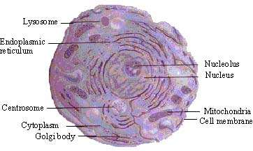
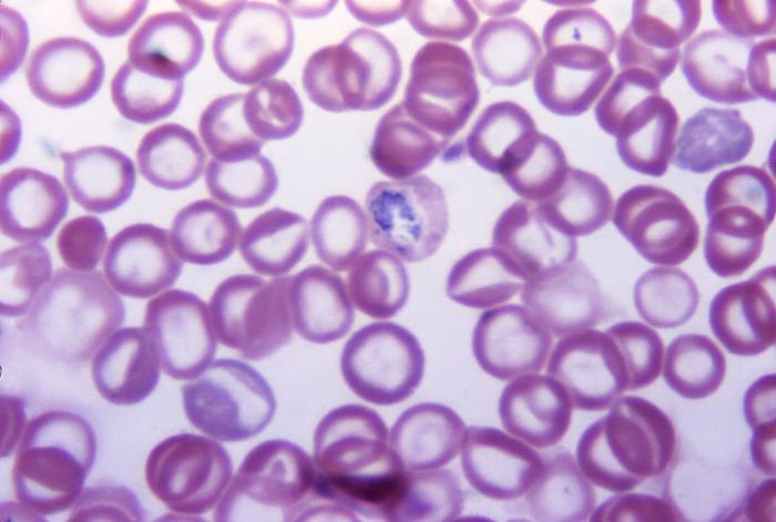
See how a generalized structure of an animal cell and plant cell look with labeled diagrams. Have the students collect their own cheek cells to prepare slides for viewing under the microscope. To compare and contrast cell structures seen using the light microscope. Smear the cotton swab on the center of the slide for 2 to 3 seconds. Learners will also identify differences between plant and animal cells. Typical animal cell center 40x. To examine the structures of actual plant and animal cells. Two slides demonstrating the cell membrane of an animal cell and the cell wall of a plant cell. To prepare and stain cells for examination with a light microscope. A microscope is required for this activity, but is not included in the. In order to view bacteria (prokayotes), which are much smaller than plant and animal cells, a different technique is used called a bacterial smear. First, to prepare an animal cell slide, start by pouring 30 ml of distilled water into a beaker. • clean sharp knife • one 2 cm cube of fresh beef or a sheep kidney purchased from a retail store.
Step 3 cover the sample with a drop of water. Take a clean cotton swab (sealed package, sterile) and gently scrape the inside of your mouth. Typical animal cell center 40x. To look at a cell close up we need a microscope. Cell is a tiny structure and functional unit of a living organism containing various parts known as organelles.
Microscopy from d32ogoqmya1dw8.cloudfront.net To compare and contrast cell structures seen using the light microscope. Add a drop of methylene blue solution on the smear. Obtain a clean microscope slide. All living organisms are made up of cells. Wet mount technique is used for preparing eukaryotic cells, such as the cells of plants and animals for the microscope. Cells have been stained to help. Prepare animal and plant cells slides. • glass microscope slides and no1 glass or plastic cover slips
How to prepare a wet mount microscope slide.
Use a razor blade to cut your specimen material into a thin, translucent slice. In order to view bacteria (prokayotes), which are much smaller than plant and animal cells, a different technique is used called a bacterial smear. Viewing animal cells under a microscope. Learners will also identify differences between plant and animal cells. Take a clean cotton swab (sealed package, sterile) and gently scrape the inside of your mouth. The typical animal cell can be seen here. Obtain a clean microscope slide. Instructions for collecting cheek cells and mounting on slide: In order to view bacteria (prokayotes), which are much smaller than plant and animal cells, a different specimen preparation technique is used called a bacterial smear. Cell is a tiny structure and functional unit of a living organism containing various parts known as organelles. 1) add one drop of food coloring to the middle of a clean slide. In this activity, learners make slides of onion cells and their own cheek cells. Both daughter cells will have.
Berbagi :
Posting Komentar
untuk "How To Prepare Animal Cell For Microscope : Fisher Science Education™ Animal Cells Basic Slide Set ... / Do not scrape your cheeks too aggressively."


Posting Komentar untuk "How To Prepare Animal Cell For Microscope : Fisher Science Education™ Animal Cells Basic Slide Set ... / Do not scrape your cheeks too aggressively."