Beranda
/ Animal Cell Diagram By Pencil : File:Simple diagram of animal cell (en).svg - Wikibooks ... : Cells are dead and have lignified secondary cell walls.
Animal Cell Diagram By Pencil : File:Simple diagram of animal cell (en).svg - Wikibooks ... : Cells are dead and have lignified secondary cell walls.
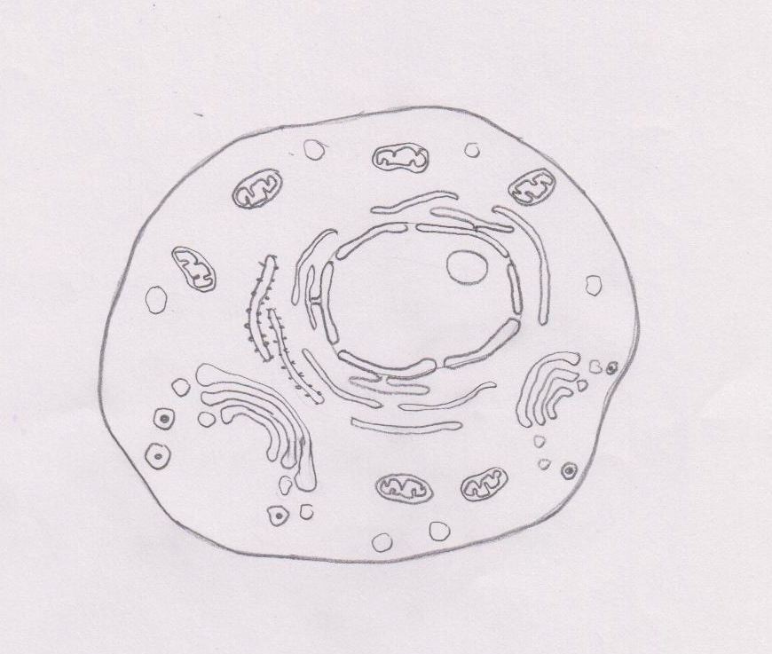
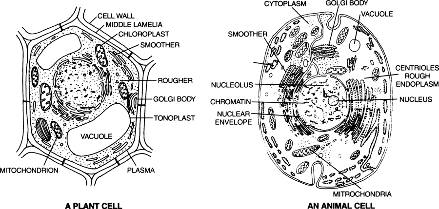
Animal Cell Diagram By Pencil : File:Simple diagram of animal cell (en).svg - Wikibooks ... : Cells are dead and have lignified secondary cell walls.. Structure found in plant cells found in animal cells cell membrane cell wall chloroplast cytoplasm nucleus vacuole 2 (b) explain why plants need chloroplasts. The onion skin cell, an example of a plant cell, generally has a rigid, rectangular shape. Cystoscopy may be recommended for any of the following conditions: If examining an animal cell, physiological saline (or contact lens solution) must be used, because if plain water is used, the cell will explode from osmotic pressure. Aug 14, 2012 · cambridge checkpoint science p1 specimen 2012 1.
The animal cell structure is the most prominent in human cheek cells. (a) complete the table by putting ticks ( ) and crosses ( ) in the correct column. Blood in the urine ();; University of cambridge international examinations cambridge checkpointscience 1113/01paper 1 for examination from 2012specimen paper 45 minutescandidates answer on the question paper.additional materials: Scientific diagrams the full lesson can be viewed by enrolling in the year 7 chemistry online course or by purchasing the year 7 chemistry lesson notes.
2.3 Eukaryotic cell - BIOLOGY4IBDP from biology4ibdp.weebly.com Aug 14, 2012 · cambridge checkpoint science p1 specimen 2012 1. In the complete animal cell centrosome, the two centrioles are arranged such that one is perpendicular to the other. Blood in the urine ();; Along this line ten light crosses ("x") are marked at intervals of about 2 cm. Unlike plant cells and bacteria, animal cells have no cell wall to structurally support them. (a) complete the table by putting ticks ( ) and crosses ( ) in the correct column. The onion skin cells were positioned beside each other (length touching length, width touching width) and formed a checkered pattern. On a clean sheet of chromatography paper with size about 12 cm by 22 cm, a light pencil line is marked to the bottom and about 1.5 cm away.
Blood in the urine ();;
Rulerread these instructions firstwrite your centre number, candidate number and name on all the work you hand in.write in dark blue or black. Aug 14, 2012 · cambridge checkpoint science p1 specimen 2012 1. On a clean sheet of chromatography paper with size about 12 cm by 22 cm, a light pencil line is marked to the bottom and about 1.5 cm away. Learning objective in this lesson we will learn the rules for drawing scientific diagrams… read more Cells are dead and have lignified secondary cell walls. Along this line ten light crosses ("x") are marked at intervals of about 2 cm. In the complete animal cell centrosome, the two centrioles are arranged such that one is perpendicular to the other. Provide the hardness of fruits like pears. Sclereids have strong walls which fill nearly the entire volume of the cell. The onion skin cells were positioned beside each other (length touching length, width touching width) and formed a checkered pattern. Loss of bladder control (incontinence) or overactive bladder; Oct 01, 2017 · other structures are found in both plant and animal cells. (although, the american urogynecologic society does not recommend that cystoscopy, urodynamics, or diagnostic renal and bladder ultrasound are part of initial diagnosis for uncomplicated overactive bladder.)
Aug 14, 2012 · cambridge checkpoint science p1 specimen 2012 1. Loss of bladder control (incontinence) or overactive bladder; Unlike plant cells and bacteria, animal cells have no cell wall to structurally support them. If examining an animal cell, physiological saline (or contact lens solution) must be used, because if plain water is used, the cell will explode from osmotic pressure. Cells are dead and have lignified secondary cell walls.
2.3 Eukaryotic cell - BIOLOGY4IBDP from biology4ibdp.weebly.com Along this line ten light crosses ("x") are marked at intervals of about 2 cm. Chloroplast has been done for you. Loss of bladder control (incontinence) or overactive bladder; The onion skin cells were positioned beside each other (length touching length, width touching width) and formed a checkered pattern. Learning objective in this lesson we will learn the rules for drawing scientific diagrams… read more On a clean sheet of chromatography paper with size about 12 cm by 22 cm, a light pencil line is marked to the bottom and about 1.5 cm away. (although, the american urogynecologic society does not recommend that cystoscopy, urodynamics, or diagnostic renal and bladder ultrasound are part of initial diagnosis for uncomplicated overactive bladder.) Cells are dead and have lignified secondary cell walls.
Cells are dead and have lignified secondary cell walls.
Oct 01, 2017 · other structures are found in both plant and animal cells. In the complete animal cell centrosome, the two centrioles are arranged such that one is perpendicular to the other. (although, the american urogynecologic society does not recommend that cystoscopy, urodynamics, or diagnostic renal and bladder ultrasound are part of initial diagnosis for uncomplicated overactive bladder.) Scientific diagrams the full lesson can be viewed by enrolling in the year 7 chemistry online course or by purchasing the year 7 chemistry lesson notes. ("u" is represents the unknown amino acid mixture). Blood in the urine ();; Structure found in plant cells found in animal cells cell membrane cell wall chloroplast cytoplasm nucleus vacuole 2 (b) explain why plants need chloroplasts. Learning objective in this lesson we will learn the rules for drawing scientific diagrams… read more The animal cell structure is the most prominent in human cheek cells. The onion skin cells were positioned beside each other (length touching length, width touching width) and formed a checkered pattern. Chloroplast has been done for you. Sclereids have strong walls which fill nearly the entire volume of the cell. On a clean sheet of chromatography paper with size about 12 cm by 22 cm, a light pencil line is marked to the bottom and about 1.5 cm away.
Unlike plant cells and bacteria, animal cells have no cell wall to structurally support them. If examining an animal cell, physiological saline (or contact lens solution) must be used, because if plain water is used, the cell will explode from osmotic pressure. Aug 14, 2012 · cambridge checkpoint science p1 specimen 2012 1. Rulerread these instructions firstwrite your centre number, candidate number and name on all the work you hand in.write in dark blue or black. Chloroplast has been done for you.
Draw diagrams of plant cell and animal cell. from Biology ... from www.zigya.com Rulerread these instructions firstwrite your centre number, candidate number and name on all the work you hand in.write in dark blue or black. Provide the hardness of fruits like pears. Oct 01, 2017 · other structures are found in both plant and animal cells. Along this line ten light crosses ("x") are marked at intervals of about 2 cm. Aug 14, 2012 · cambridge checkpoint science p1 specimen 2012 1. In the complete animal cell centrosome, the two centrioles are arranged such that one is perpendicular to the other. Chloroplast has been done for you. ("u" is represents the unknown amino acid mixture).
The animal cell structure is the most prominent in human cheek cells.
Cystoscopy may be recommended for any of the following conditions: (although, the american urogynecologic society does not recommend that cystoscopy, urodynamics, or diagnostic renal and bladder ultrasound are part of initial diagnosis for uncomplicated overactive bladder.) In the complete animal cell centrosome, the two centrioles are arranged such that one is perpendicular to the other. Chloroplast has been done for you. Provide the hardness of fruits like pears. Loss of bladder control (incontinence) or overactive bladder; Structure found in plant cells found in animal cells cell membrane cell wall chloroplast cytoplasm nucleus vacuole 2 (b) explain why plants need chloroplasts. The onion skin cells were positioned beside each other (length touching length, width touching width) and formed a checkered pattern. University of cambridge international examinations cambridge checkpointscience 1113/01paper 1 for examination from 2012specimen paper 45 minutescandidates answer on the question paper.additional materials: Rulerread these instructions firstwrite your centre number, candidate number and name on all the work you hand in.write in dark blue or black. On a clean sheet of chromatography paper with size about 12 cm by 22 cm, a light pencil line is marked to the bottom and about 1.5 cm away. Oct 01, 2017 · other structures are found in both plant and animal cells. If examining an animal cell, physiological saline (or contact lens solution) must be used, because if plain water is used, the cell will explode from osmotic pressure.
Berbagi :
Posting Komentar
untuk "Animal Cell Diagram By Pencil : File:Simple diagram of animal cell (en).svg - Wikibooks ... : Cells are dead and have lignified secondary cell walls."

Posting Komentar untuk "Animal Cell Diagram By Pencil : File:Simple diagram of animal cell (en).svg - Wikibooks ... : Cells are dead and have lignified secondary cell walls."